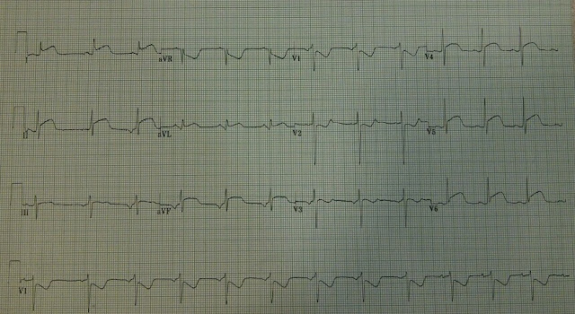
This patient presented to an outside hospital for palpitations following the use of a routine anti-asthma medication. The patient reported chest discomfort and some associated shortness of breath shortly after medication administration. The patient's vital signs remain stable, and her lung sounds were clear. The remainder of the physical examination was unremarkable. An emergency physician obtained a routine ECG. The patient's vital signs were stable and the physical examination was unremarkable.
The patient was transferred to a tertiary care facility for cardiac catheterization.
1. What's your interpretation of the 12 lead?
2. What are some diagnostic considerations?
2. What are some diagnostic considerations?
12 lead ECG Interpretation
Sinus rhythm, rate approximately 70, diffuse ST segment elevation
There is a baseline sinus rhythm. Widespread ST segment elevation is present in leads I, aVL, II, III, aVF, and in the anterior-lateral precordial leads. The R waves and ST segment depression in leads V1 and V2 are consistent with posterior wall ischemia. The ST segments themselves are mostly convex. The convexity of the ST segment is also suggestive of ischemia. Infarction of the heart's anterior, inferior, lateral, and posterior walls could conceivably produce the diffuse ST segment changes seen in this ECG.
12 lead ECG Discussion and Case Resolution
There are several considerations to bear in mind when looking at diffuse ST segment elevations:
1. Massive myocardial infarction
2. Pericarditis
3. Ventricular wall aneurysm
4. Coronary vasospasm
The patient's clinical picture (and overall well appearance) is not consistent with the diagnosis of a massive myocardial infarction.
The ST elevations in pericarditis are usually more CONCAVE in appearance. Reciprocal changes are never associated with pericarditis. PR segment depression is also present in pericarditis.
Ventricular wall aneurysm usually presents electrocardiographically with Q waves and diffuse ST segment elevations. There may be a loss of R wave progression across the precordial leads.
There was no lesion amenable to intervention at the time of angiography. The patient's echocardiogram revealed a preserved ejection fraction with some mild, but diffuse, hypokinesis. The patient was diagnosed with coronary artery vasospasm.

No comments:
Post a Comment